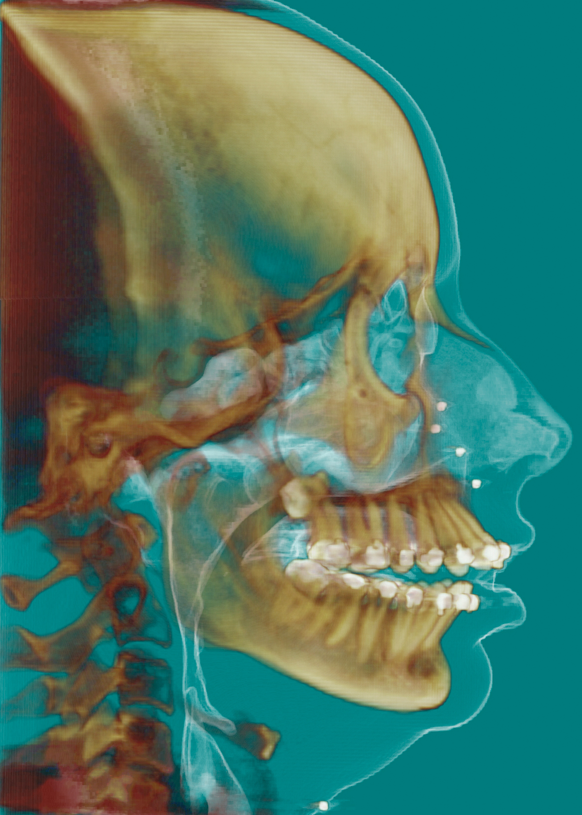


DOLPHIN IMAGING WINDOWS
DCMprinter receives images from DICOM modalities and prints them on a regular Windows paper printer. That means you can send DICOM images stored on your OsiriX HD database to any DICOM nodes, including another OsiriX HD iOS device, OsiriX for macOS, a DICOM viewer, or a PACS server. Custom URL Scheme dicom-shot:// Notes - Requires a Wi-Fi environment to the clinic.
DOLPHIN IMAGING SOFTWARE
If the software did not start review section 2.

I know, this function dont send information the patient, study and series. SendToPACS converts non-DICOM images, video and signal files into DICOM format and sends DICOM format files to a PACS server/VNA or DICOM workstation. DICOM (Digital Imaging and Communications in Medicine) Staff Contact: Carolyn Hull. Convert and send Word files, PDFs, reports, emails, and images with the PACS Scan Virtual Printer, a printer selection that appears DICOM Processing and Segmentation in Python. DICOM (Digital Imaging and Communications in Medicine) is an application layer network protocol for the transmission of medical images, waveforms and accompanying information. C-STORE is the operation that allow sending a DICOM file to a remote DICOM server. I am using a ClearCanas PACS server and have access to the web GUI. There is a need for a separate PACS server that supports the DICOM communication protocol. We have made an effort to keep these pages as clearly arranged as possible. Digital Imaging and Communications in Medicine (DICOM) is an international standard used for medical images such as X-rays, MRIs, ultrasounds, and other medical imaging modalities. description: Pictures of follow-up,Pictures of observation,Pictures preoperative,Pictures intraoperative,Pictures postoperative: no: A: Comma-separated list of study description elements. DICOM acts as both a file format and the international communication standard through which PACS transfers medical image data, while PACS drives the DICOM workflow. These findings indicate that Version 8.0 of Dolphin Imaging Software needs to be re-assessed for software errors that may result in clinically significant miscalculations, and to facilitate compensation of radiographic magnification when using linear measurements.Dicom send The DICOM image used in this tutorial is from the NIH Chest X-ray dataset. The investigation revealed the impact of radiographic magnification when used in an uncalibrated system.
DOLPHIN IMAGING MANUAL
However, systematic error in the software's calculation of LAFH% resulted in measurements 4% larger than manual techniques, a difference which is clinically significant.Ĭomparison of actual outcome and software generated prediction for 26 orthognathic cases demonstrated clinically significant differences for all measurements (ρc 0.32 for ANB to 0.91 for LIMd P < 0.05). Comparing the standard deviations of the differences, manual tracing proving more reliable for SNA (1.36° manually, 2.07° digitally), SNB (1.19° and 1.69°), SNMx (1.39° and 2.66°), and MxMd (1.77° and 2.26°), and Dolphin digital tracing more reliable for UIMx (3.49° digitally and 3.97° manually) and LIMd (2.90° and 3.04°). Method error (reliability) using duplicate measurements for each method, and comparison of both techniques (reproducibility), were investigated using alternative statistical methods, Bland and Altman (1986) and Lin's Correlation of Concordance (1989).Įach technique was significantly reliable at the 95% level (method error). Sixty lateral cephalograms were evaluated by two methods: manual tracing and indirect digitization using Dolphin Imaging Software (Version 8.0). In addition, orthognathic prediction was compared with actual outcomes. The purpose of this study was to examine and compare the reproducibility and reliability of digitization using Dolphin Imaging Software (Version 8.0) with traditional manual techniques.


 0 kommentar(er)
